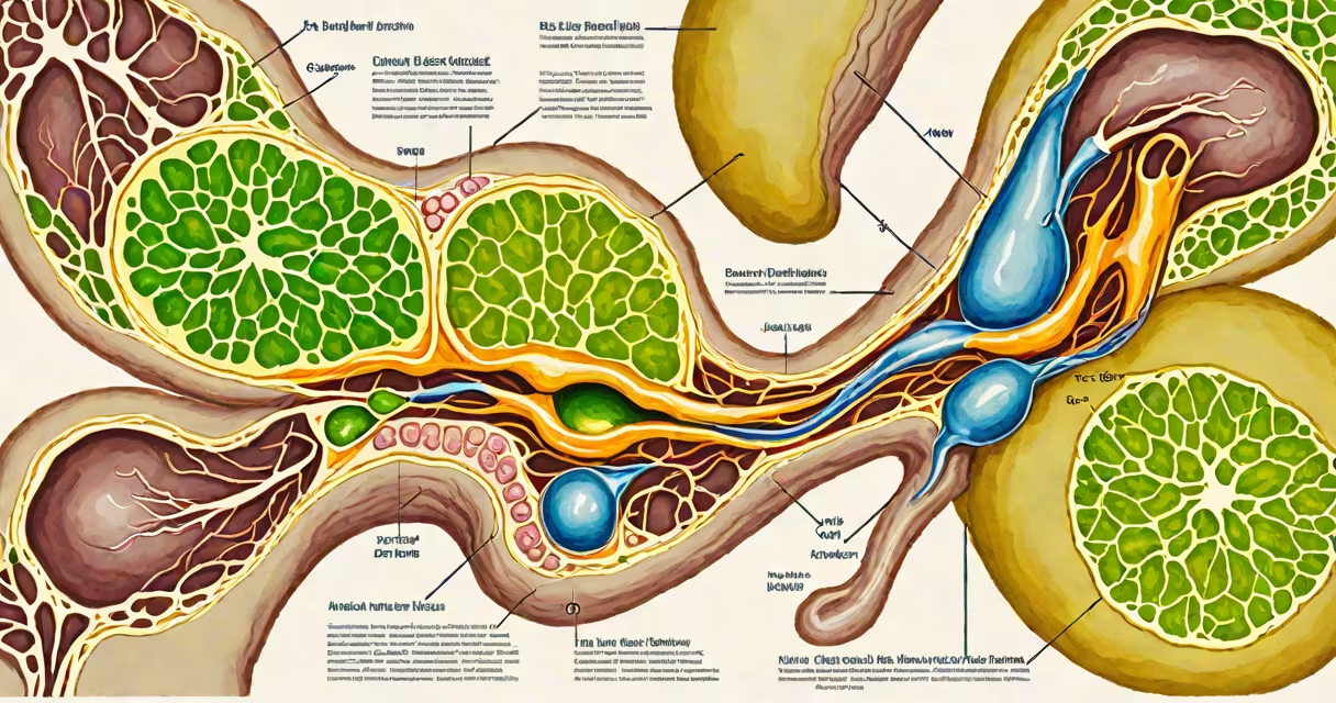
Biliary dilatation occurs when the bile ducts widen due to blockage or pressure buildup, often indicating obstruction in the biliary system. This condition arises from physical blockages, inflammation, or structural abnormalities affecting bile flow.
When bile cannot drain freely from the liver through the common bile duct into the duodenum, pressure increases upstream, causing dilation. The most frequent cause is choledocholithiasis-gallstones lodged in the common bile duct. Other contributors include malignant tumors, benign strictures, and inflammatory conditions such as primary sclerosing cholangitis.
Imaging reveals dilatation as widened ducts visible on ultrasound, CT, or MRCP. A duct diameter exceeding 6 mm in the common bile duct is considered abnormal. Persistent dilatation signals underlying pathology requiring intervention.
Bile produced in the liver flows through intrahepatic ducts into the common hepatic duct, then merges with the cystic duct from the gallbladder to form the common bile duct. This duct empties into the duodenum via the ampulla of Vater. Any obstruction along this path leads to upstream dilation.
Dilation begins distal to the site of blockage. For example, a stone in the distal common bile duct causes the entire duct and intrahepatic branches to enlarge. The liver's ability to secrete bile continues, but drainage fails, leading to increased pressure and duct expansion.
Chronic obstruction results in duct wall thinning and fibrosis. Acute cases may trigger cholangitis-life-threatening infection with fever, right upper quadrant pain, and elevated bilirubin. Early recognition prevents complications like liver abscess or sepsis.
The most common causes fall into four categories: mechanical obstruction, inflammatory disease, congenital anomalies, and neoplastic processes.
| Category | Specific Cause | Prevalence in Studies | Key Imaging Finding |
|---|---|---|---|
| Mechanical Obstruction | Gallstones in common bile duct | 60-70% of obstructive cases | High-density stone with acoustic shadow on ultrasound |
| Inflammatory Disease | Primary sclerosing cholangitis | 1-2% of chronic liver disease patients | Beaded narrowing and segmental strictures on MRCP |
| Neoplastic | Pancreatic head adenocarcinoma | Responsible for 15-20% of extrahepatic obstructions | Strictures with abrupt cutoff on cross-sectional imaging |
| Congenital | Choledochal cyst | Rare; seen in 1 in 15,000 births | Fluid-filled dilation of the extrahepatic ducts |
Among these, choledocholithiasis remains the leading cause. A 2021 study in *Gastroenterology* found that 68% of patients with biliary dilatation on ultrasound had stones confirmed during endoscopic retrograde cholangiopancreatography (ERCP).
Primary sclerosing cholangitis (PSC) presents in younger adults, often with inflammatory bowel disease. Ducts appear irregular and narrowed on MRCP, mimicking malignancy. Biopsy is sometimes needed to differentiate PSC from cholangiocarcinoma.
Neoplasms such as pancreatic cancer compress the common bile duct. The classic sign is a "double-duct sign" on CT: dilated common bile duct and pancreatic duct due to tumor mass near the ampulla.
Some assume biliary dilatation always indicates malignancy. That's incorrect. Gallstones are far more common than tumors in obstructive cases. Over 60% of biliary dilatation episodes stem from benign causes.
Another error is equating mild dilation with clinical significance. A duct measuring 7 mm may be normal in older adults due to age-related changes. However, a sudden increase from 4 mm to 8 mm over weeks suggests progression and warrants investigation.
A non-obvious point: biliary dilatation can occur without jaundice. Some patients present with abdominal pain or fever alone. Bilirubin levels remain normal if the obstruction is partial or intermittent. This delays diagnosis until symptoms worsen.
A 62-year-old woman presented with severe right upper quadrant pain and fever. Labs showed elevated alkaline phosphatase (1,200 U/L) and direct bilirubin (3.2 mg/dL). Ultrasound revealed a 12-mm dilated common bile duct with a 10-mm hyperechoic focus with posterior shadowing.
These findings indicated choledocholithiasis. She underwent ERCP the next day. A stone was extracted, and her symptoms resolved within 24 hours. Follow-up ultrasound confirmed normalization of duct size.
This case illustrates that dilatation is a red flag, not a diagnosis. The cause must be identified using imaging and invasive procedures when needed. MRCP is first-line for non-invasive evaluation, while ERCP offers both diagnosis and treatment.
Understanding biliary dilatation causes enables timely management. Gallstones, strictures, tumors, and inflammatory diseases are the core culprits. Accurate diagnosis requires integrating imaging, lab values, and clinical context. Ignoring subtle signs risks missing treatable conditions.