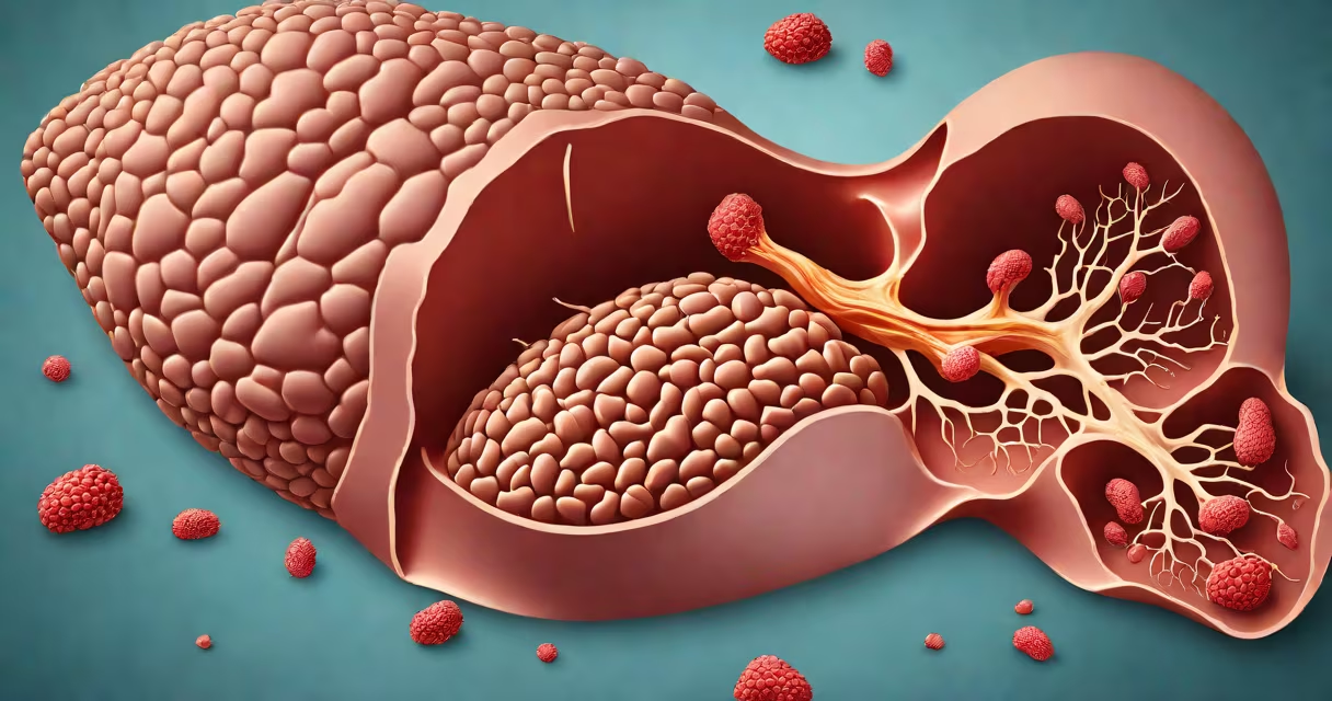
Liver cirrhosis is irreversible scarring of the liver tissue resulting from chronic injury, leading to loss of function and altered blood flow. It develops over years due to persistent inflammation and fibrosis, replacing healthy liver cells with nonfunctional scar tissue.
Cirrhosis occurs when the liver repeatedly repairs itself after damage, forming dense fibrous bands that disrupt normal structure. These changes impair detoxification, protein synthesis, and bile production. The condition progresses through stages-from compensated to decompensated-with increasing risk of life-threatening complications like variceal bleeding or hepatic encephalopathy.
According to the American Association for the Study of Liver Diseases (AASLD), cirrhosis affects approximately 1 in 400 adults in the United States, with alcohol-related liver disease and non-alcoholic fatty liver disease (NAFLD) being primary drivers. Early diagnosis remains critical because symptoms often appear only after significant damage has occurred.
The process begins with ongoing injury-such as excessive alcohol consumption, viral hepatitis B or C, or fat accumulation in NAFLD-that triggers chronic inflammation. Immune cells release signaling molecules that activate hepatic stellate cells, which then produce collagen and other extracellular matrix proteins.
Over time, this leads to fibrosis-excessive deposition of connective tissue. As fibrosis spreads, it forms regenerative nodules surrounded by scar tissue. These nodules disrupt blood flow through the liver, causing portal hypertension, where pressure builds in the portal vein system.
A 2021 study published in *Gastroenterology* found that patients with sustained ALT levels above 50 U/L and AST/ALT ratios greater than 1 had higher odds of progressing to cirrhosis within five years. This highlights the role of enzyme patterns in tracking disease severity.
Cirrhosis is classified by etiology-the root cause. The most common types include:
| Primary Cause | Prevalence in U.S. Cirrhosis Cases | Key Risk Factors |
|---|---|---|
| Alcohol-associated liver disease (ALD) | 30-40% | Chronic heavy drinking (>60g/day for men, >40g/day for women) |
| Non-alcoholic fatty liver disease (NAFLD) | 30-35% | Obesity, insulin resistance, type 2 diabetes |
| Hepatitis B and C | 10-15% | Unprotected sex, IV drug use, blood exposure |
| Autoimmune hepatitis | 5-10% | Genetic predisposition, female predominance |
| Hereditary conditions (e.g., hemochromatosis, Wilson's disease) | <5% | Genetic mutations affecting iron or copper metabolism |
Each type follows a similar path: initial injury → inflammation → fibrosis → nodule formation → functional decline. However, progression rates differ. For example, individuals with untreated hepatitis C have a 15% risk of developing cirrhosis within 20 years, according to the CDC.
One lesser-known fact: not all cirrhosis is symptomatic. Up to 50% of people with compensated cirrhosis report no noticeable symptoms until imaging or lab tests reveal abnormalities. This silent phase increases the chance of sudden decompensation.
Cirrhosis is often mistaken for fatty liver or hepatitis. Fatty liver involves fat buildup without significant scarring. Hepatitis refers to inflammation, which may resolve without lasting damage. Only when inflammation persists do fibrotic changes begin-and only after years do they reach the threshold of cirrhosis.
Another misconception is that cirrhosis is always fatal. While it reduces life expectancy, many patients live for decades with stable, compensated disease. A 2020 cohort study in *The Lancet Gastroenterology & Hepatology* showed that 70% of patients with compensated cirrhosis survived at least 10 years with lifestyle modification and medical management.
It's also incorrect to assume that stopping alcohol use reverses cirrhosis. Abstinence halts further damage and may improve liver function, but existing scar tissue does not regenerate. The goal becomes preventing progression and managing complications.
Consider a 54-year-old man with a 20-year history of daily alcohol intake. He presents with fatigue and mild jaundice. Lab results show elevated AST (120 U/L), ALT (80 U/L), and a serum albumin of 3.2 g/dL. An ultrasound reveals a nodular liver surface and splenomegaly.
He undergoes a FibroScan, which measures liver stiffness at 28 kPa-above the threshold of 12.5 kPa indicating cirrhosis. A biopsy confirms bridging fibrosis and regenerative nodules. His Child-Pugh score is 6 (Class A), indicating compensated cirrhosis.
After referral to a hepatologist, he stops drinking, receives vitamin supplementation, and begins monitoring for esophageal varices via endoscopy every two years. His condition stabilizes over the next three years with no episodes of ascites or confusion.
This case illustrates that early diagnosis and intervention can halt progression-even in advanced cases. The patient's survival depends less on reversing scarring than on controlling risk factors and detecting complications early.
Patients should consult a specialist if they have persistent fatigue, unexplained weight loss, swelling in the legs, or dark urine. Routine screening is recommended for those with known risk factors, including annual liver function tests and ultrasound for high-risk groups.
Specialists who manage cirrhosis include hepatologists, gastroenterologists with subspecialty training, and transplant surgeons. General practitioners often initiate evaluation but refer to specialists for staging, treatment planning, and complication management.
Liver cirrhosis is permanent scarring of the liver due to prolonged injury. It results from conditions like alcohol abuse, NAFLD, and viral hepatitis. The disease progresses through stages, with early phases often asymptomatic. Diagnosis relies on imaging, lab tests, and sometimes biopsy. Treatment focuses on halting progression, managing complications, and identifying candidates for transplantation.
Understanding what is liver cirrhosis enables earlier intervention and better outcomes. With proper care, many patients maintain quality of life for years. Awareness of risk factors and regular monitoring remain central to reducing preventable deaths.